Are you developing antibody-drug conjugates (ADCs)?
Are you seeking a reliable and standardized method to evaluate antibody internalization?
We are proud to introduce the DT3C Protein (Cat. No. AME100004) — a novel recombinant fusion protein designed to simulate ADC mechanisms and provide a fast, universal, and functional solution for antibody internalization assessment.
1. What is DT3C?
DT3C is a recombinant fusion protein combining Diphtheria Toxin Fragment A and the Streptococcus Protein G Fc-binding domains (C1–C3).
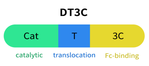
Fig 1. The struture of DT3C protein.
1.1 Diphtheria Toxin Fragment A (DT-A)
DT-A is the active domain of diphtheria toxin with ADP-ribosyltransferase activity, responsible for its cytotoxic effect.
Mechanism of action: Once inside the cell, DT-A catalyzes the transfer of an ADP-ribose group from NAD⁺ to elongation factor-2 (EF-2). This modification inactivates EF-2, blocking the translocation step in translation, ultimately inhibiting protein synthesis and inducing cell death.
In DT3C, the function of DT-A is:
- Dependency on internalization: DT-A alone cannot enter cells; its activity depends on antibody-mediated uptake.
- Mimicking ADC payload release: Once internalized, DT-A exerts its toxicity in the cytosol, simulating ADC intracellular action.
1.2 Translocation Domain
The translocation domain, derived from the central “T domain” of diphtheria toxin, enables escape from endosomal compartments into the cytosol.
In acidic environments, it undergoes conformational changes to deliver the toxic payload across the membrane. This mechanism is common in bacterial toxins like diphtheria and anthrax toxin.
1.3 Streptococcus Protein G Fc-binding Domains (C1–C3)
The C1–C3 domains of Streptococcus Protein G bind the Fc region of IgG antibodies with high affinity, without interfering with antigen binding (Fab region).
In DT3C, these domains:
- Mediate non-covalent binding to IgG Fc from various species (human, mouse, rabbit, etc.);
- Enable the formation of mAb–DT3C complexes;
- Support antibody-guided targeting and internalization into specific cells;
DT3C binds to the Fc portion of IgG antibodies, forming a mAb–DT3C complex. Upon target antigen recognition, the complex is internalized by the cell.
Inside the cell, DT-A is released, mimicking ADC payload release and cytotoxic function.
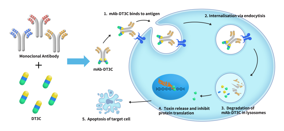
Fig 2. Working principle diagram of DT3C protein.
2. Product Advantages
- No chemical conjugation needed
- Broad compatibility with multiple IgG species and subclasses
- Rapid evaluation of internalization and functional toxicity
- Suitable for high-throughput screening and diverse cell platforms
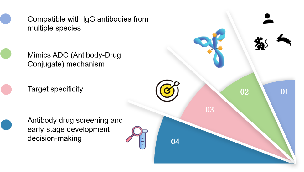
Fig 3. The advantages of DT3C protein.
3. Case Study
DiTag™ DT3C IgG Labeling Reagent (AME100004)
- SDS-PAGE Purity Validation
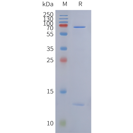
Fig 4. DT3C Protein on SDS-PAGE under reducing condition.
- Antibody Internalization Assay
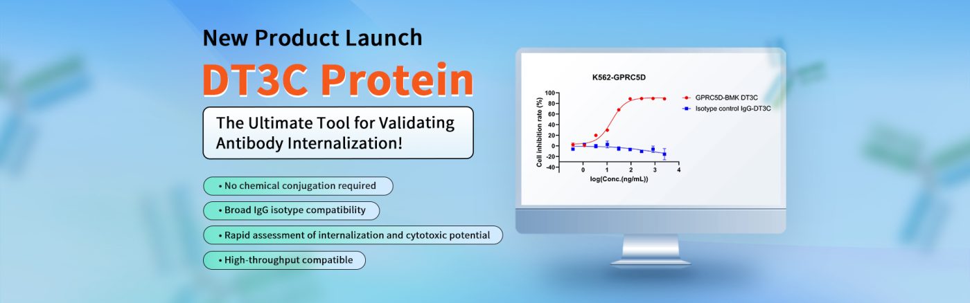
CCK-8 assay on K562-GPRC5D cells to assess cytotoxicity. The ADC control BMK showed an IC50 of 15.55 ng/mL, indicating effective internalization and target-specific action.
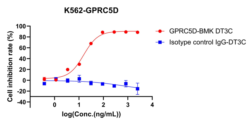
Fig 5. Cell inhibition rate of K562-GPRC5D detected by CCK8 method. The IC50 of GPRC5D ADC BMK (BME100187) is 15.55ng/ml, indicating specific internalization.
4. FAQ – Quick Q&A
| Question | Answer |
|---|---|
| Which antibodies can DT3C bind? | IgG from human, mouse, rabbit, and more |
| Is chemical conjugation needed? | No. DT3C binds Fc non-covalently |
| Is DT3C suitable for live-cell studies? | Yes, ideal for internalization & cytotoxicity assays |
| What detection methods are supported? | Fluorescence, flow cytometry, imaging, CCK-8, etc. |
| How should it be stored/used? | Store at –80°C, avoid freeze-thaw, incubate with antibody at recommended ratio |